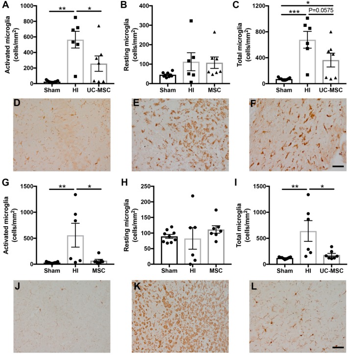
Figure 4.
Microglial activation is increased following neonatal HI brain injury and reduced by UC-MSC treatment. Iba-1 immunohistochemistry was performed to identify microglia in the hippocampus, somatosensory cortex and periventricular striatum region of the brain. (A) Activated microglia, (B) resting microglia and (C) total microglia in the hippocampus. Representative images of Iba-1 staining in the hippocampus for (D) sham, (E) HI and (F) MSC treatment; (G) Activated microglia, (H) resting microglia, and (I) total microglia in the somatosensory cortex. Representative images of Iba-1 staining in the somatosensory cortex for (J) sham, (K) HI and (L) MSC treatment. All images were taken at 400× magnification, scale bar = 500 μm (n = 6–9 rats per group, * p < 0.05, ** p < 0.01, *** p < 0.001).

