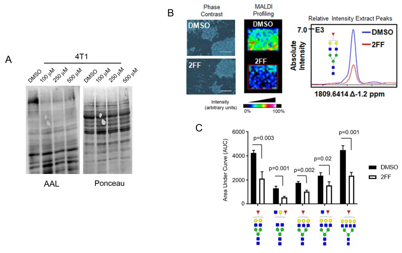
Figure 1.
2-deoxy-2-fluoro-L-fucose (2FF) suppress fucosylation in 4T1 cells. (A) Decreased fucose detected by Aleuria aurantia lectin (AAL) blotting of 4T1 cell lysates treated with vehicle (DMSO) or increasing concentration (100–500 μM) of 2FF, a fucosylation inhibitor. Ponceau S stain was used to show total protein loading. (B) MALDI-IMS of 4T1 cells grown in tissue chambered slide and treated with DMSO or with 500 μM 2FF. Phase contrast (left panel) depicts cells from DMSO or 2FF chambers used in MALDI profiling experiments (middle panel). Scale bar = 400 µm The graph showing absolute intensity is shown (right panel). (C) The area under the curve (AUC) of normalized total ion count (TIC) was calculated and compared between DMSO control and 2FF treated samples. Glycan nomenclature: Blue square as GlcNAc, yellow circle as galactose, green circle as mannose, red triangle as fucose.

