Figure 5.
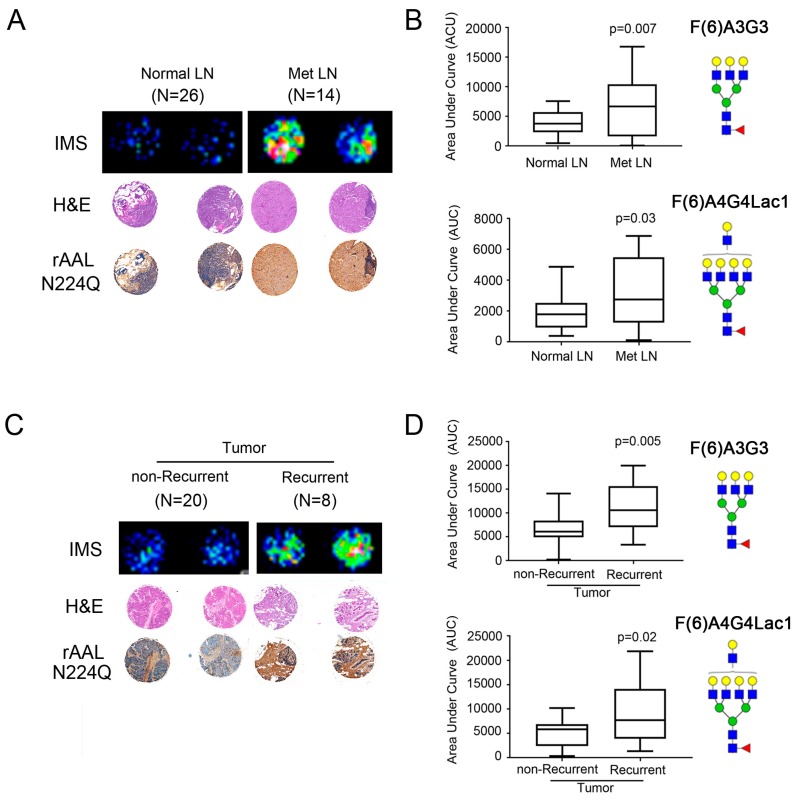
Increased fucosylation in metastatic lymph nodes (met LN) and tumors from recurrent patients. (A) A representative comparison of a patient with normal LN and met LN (n = 33 patients) showed the increased distribution of fucosylated glycan (F(6)A4G4Lac1 shown here). The H&E stain was used to show the tumor region. Increased core fucosylation in met LN and tumors from patients with recurrence confirmed with lectin staining using rAALN224Q, a lectin with enhanced binding to core-fucosylated structures. TMA cores were imaged at 50× magnification. (B) Statistically significant higher core fucosylated tri-antennary glycans (F(6)A3G3) and core fucosylated tetra-antennary with one lactosamine arm (F(6)A4G4Lac1) in the met LN samples (p < 0.05). (C) A representative comparison of a patient with non-recurrent and recurrent tumors (n = 33 patients) showed the increased distribution of fucosylated glycan by MALDI-IMS, with H&E staining, and confirmed with lectin staining using rAALN224Q. TMA cores were imaged at 50× magnification. (D) Statistically significant higher core fucosylated tri-antennary glycans (F(6)A3G3) and core fucosylated tetra-antennary with one lactosamine arm (F(6)A4G4Lac1) in the met LN samples (p < 0.05).

