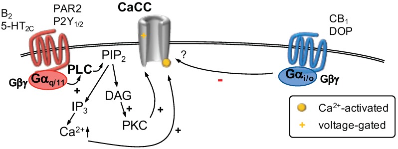
Figure 4.
Calcium-activated Cl channels (CaCC) are gated by increasing concentrations of cytosolic Ca and influenced by membrane voltage. The source of Ca may be Ca-permeable ion channels located in proximity to CaCCs (not shown) or Ca released from intracellular stores. Stimulation of G-coupled receptors (left) activates phospholipase C (PLC). The subsequent hydrolysis of phosphatitylinositol 1,4, bisphosphate (PIP) forms inositiol 1,4,5 trisphosphate (IP) and diacylglycerol (DAG). DAG activates protein kinase C (PKC) which influences CaCC function. IP binds to IP receptors located in the membrane of the endoplasmic reticulum (ER) which causes Ca release from the ER. Activation of G-coupled receptors (right) was shown to decrease CaCC currents via an unknown mechanism.

