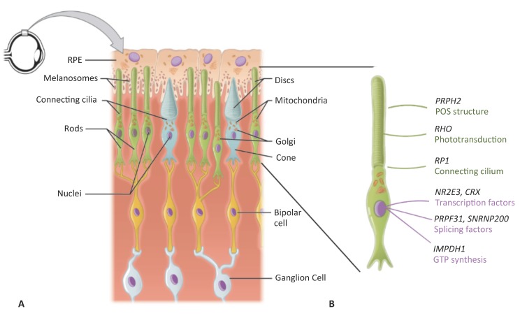
Figure 1.
Schematic representation of the retina. (A) The mono-layered retinal pigment epithelium (RPE) is located on the posterior side of the retina. It contains apically located melanosomes that provide its pigmentation. The RPE is in close contact with the outer segments of the rod (in green) and cone (in blue) photoreceptors. Each outer segment, which contains the lipid discs important for phototransduction, is connected to the cell body of the photoreceptor by a connecting cilium. On the anterior side, the photoreceptors synapse with bipolar cells (in yellow), which in turn synapse with the retinal ganglion cells (in grey). (B) Higher magnification of a rod photoreceptor shown in A), depicting the characteristic rod structure and the site of action of the proteins encoded by the genes reviewed in this article. Modified from Wikimedia Commons (author OpenStax college). License to reproduce: https://creativecommons.org/licenses/by/3.0/legalcode.

