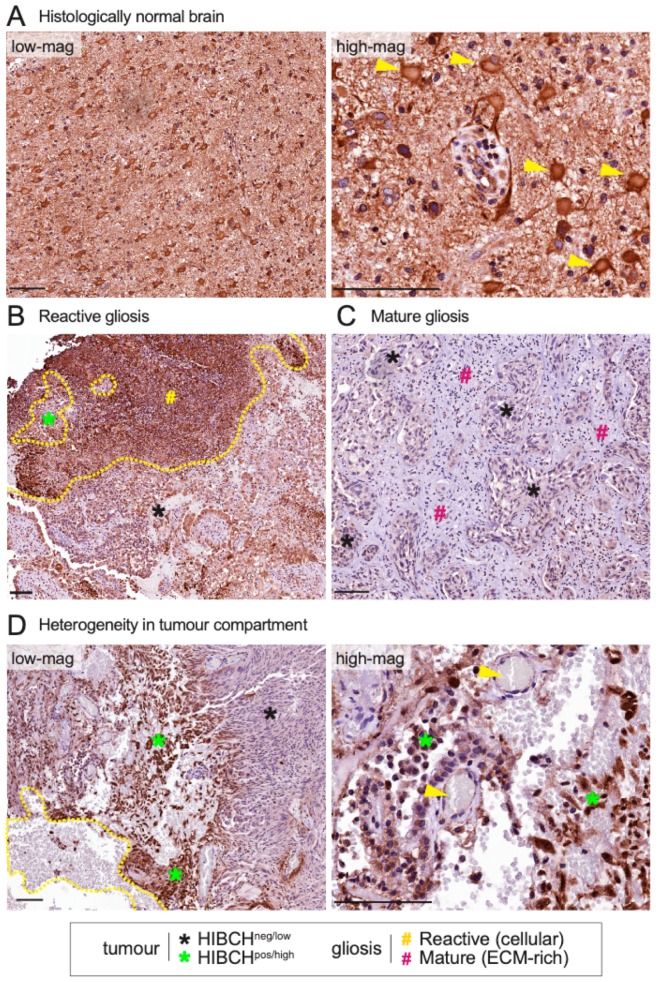
Figure 5.
IHC analysis of HIBCH expression in human triple-negative breast cancer brain metastases —key observations from five cases. (A) Representative staining in histologically normal brain tissue, at low and high magnification, showing large neurons with strong cytoplasmic expression (yellow arrows) (B) Representative strong HIBCH staining in reactive gliosis. The yellow line demarcates gliotic from metastatic tissue. (C) Extracellular matrix (ECM)-rich gliosis surrounding HIBCH-negative tumour cells. (D) Heterogeneity in the tumour compartment. Yellow line encapsulates haemorrhagic tissue and the enlargement shows small cerebral blood vessels (yellow arrows). Scale bars: 100 μm.

