Figure 1.
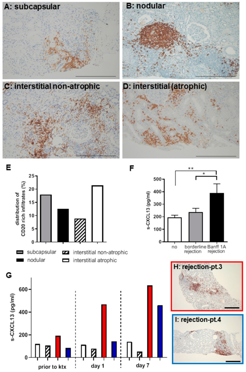
CD20+ cells were detected as part of inflammatory infiltrates in patient biopsies with TCMR (A–D, bar: 100 μm)) in subcapsular, tubular-interstitial (atrophic and non-atrophic) areas as well as in nodular infiltrates. In the non-rejection state, no CD20 positivity is detectable (data not shown). CD20+ cells were quantified in 67 randomly selected human biopsies with TCMR and graded as B-cell rich (>30 CD20-positive cells/hpf). In subcapsular infiltrates, 17.9%, in interstitial-nodular infiltrates, 12.5%, and in interstitial/atrophic areas, 21.4% were B-cell rich. The Banff-relevant interstitial non-atrophic areas contained 8.9% B-cell rich infiltrates (E). Serum CXCL13 levels are increased in patients with TCMR (Banff1a) compared to patients with borderline or no rejection (** p = 0.01; * p = 0.05) (F). Four patients had CXCL13 measurements during the first week after ktx (G). All had low levels of CXCL13 prior to ktx and two patients developed a relevant increase of CXCL13 levels up to day 7. Biopsy revealed a rejection with B-cell rich infiltrates (H,I, bar: 200 μm).

