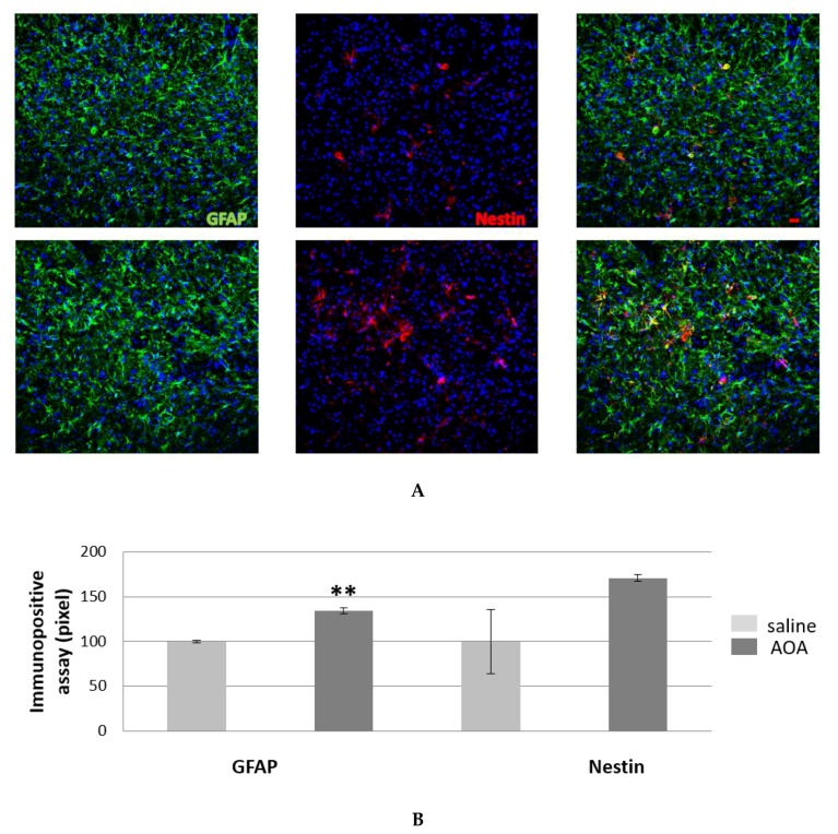
Figure 3.
GFAP and nestin positive cells in the spinal cord of SOD1G93A saline- and AOA-treated mice. The double immunofluorescence of GFAP and nestin reveals an astrocyte increase in AOA-treated mice (bottom panel, (A) compared to those treated with saline (upper panel, A), also showed from immunopositive assay (B). The mean of the pixels was quantified in three fields of the ventral horn spinal cord in AOA-treated mice (n = 3) and saline-treated mice (n = 3), data are presented as a percentage of normalised to saline values as the mean ± SEM. The values were compared by using Student’s t-test with **p < 0.01 (B). Scale bar = 20 microns.

