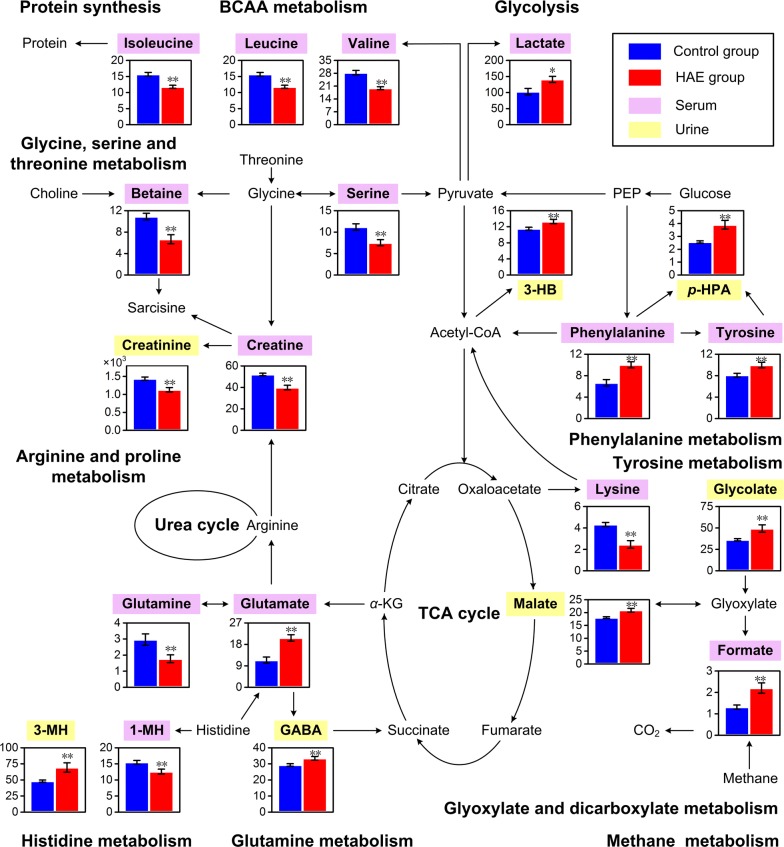
Fig. 7.
Mapping of differential metabolites induced by HAE onto metabolic pathways. Each bar graph represents one metabolite in relative concentration (mean ± standard error) of control (blue) and HAE (red) groups. *Indicates P < 0.05 statistical significance relative to control group; **Indicates P < 0.01 statistical significance relative to control group

