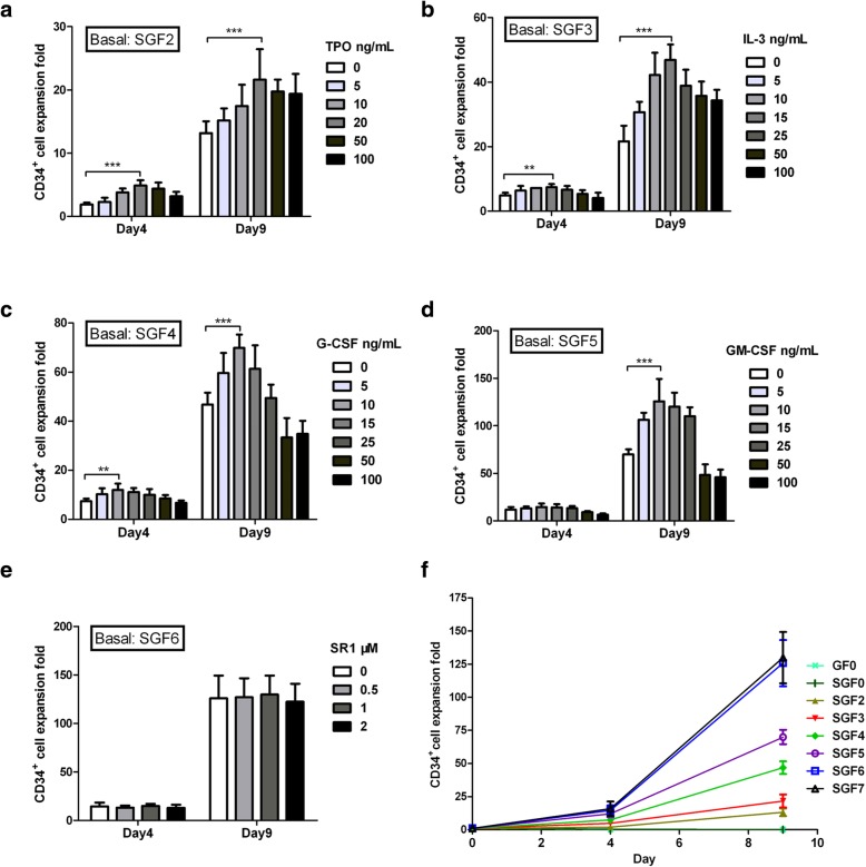
Fig. 1.
Effect of different medium conditions on HSC expansion ex vivo. a CD34+ cell expansion with TPO concentrations ranging from 0 to 100 ng/mL in SGF2 medium (IMDM supplemented with 200 ng/mL SCF, 200 ng/mL Flt-3 L, and nutrition supplements). b CD34+ cell expansion with IL-3 concentrations ranging from 0 to 100 ng/mL in SGF3 medium (SGF2 with 20 ng/mL TPO). c CD34+ cell expansion with G-CSF concentrations ranging from 0 to 100 ng/mL in SGF4 medium (SGF3 with 15 ng/mL IL-3). d CD34+ cell expansion with GM-CSF concentrations ranging from 0 to 100 ng/mL in SGF5 medium (SGF4 with 10 ng/mL G-CSF). e CD34+ cell expansion with SR1 concentrations ranging from 0 to 2 μM in SGF6 medium (SGF5 with 10 ng/mL GM-CSF). f A summary of CD34+ cell expansion in media with different growth factor combinations. SGF7 is SGF6 supplemented with 1 μM SR1. Expansion was calculated as fold increase (after/before expansion) in cell counts on each day (days 0, 4, and 9). Data are shown as mean ± SD, n = 6. **p < 0.01, ***p < 0.001; one-way ANOVA followed by Dunnett’s multiple comparison test

