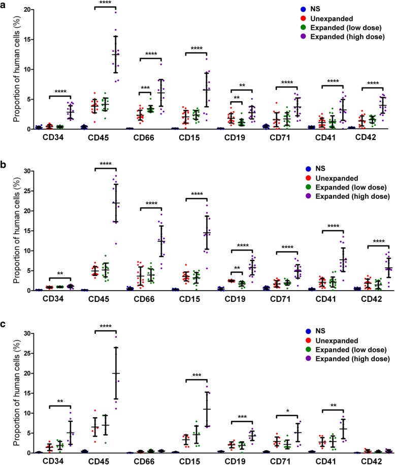
Fig. 3.
Functional assessment of human cells in PB and BM of NOD/SCID mice transplanted with various types of human HSCs. Three weeks (a) and eight weeks (b) after intravenous human CD34+ transplantation in mice, the presence of human cells was analyzed in the PB of mice transplanted with unexpanded human HSCs, low-dose expanded HSCs, or high-dose expanded HSCs. Normal saline (NS) was injected as the vehicle control. Data are shown as mean ± SD, n = 16. c At week 8 post-transplantation, engrafted human cells were detected in the BM by flow cytometry. The percentages of cells expressing human hematologic-lineage markers shown are calculated on the total (mouse plus human) cell population from mouse PB or BM. Data are shown as mean ± SD, * p < 0.05, **p < 0.01, ***p < 0.001, ****p < 0.0001; one-way ANOVA followed by Dunnett’s multiple comparison test

