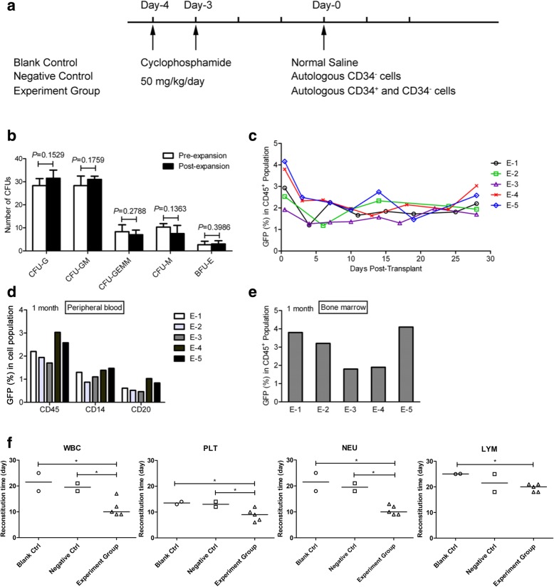
Fig. 5.
Autologus transplantation in a nonhuman primate model. a Scheme for autologus transplantation in a nonhuman primate model. CTX treatment was administered on days − 4 and − 3, and transplantation was performed on day 0. b Types of CFU colonies formed from fresh isolated CD34+ cells (pre-expansion) and 9-day HEM-expanded cells (Post-expansion). Data are shown as mean ± SD, n = 4. c Percentage of GFP+ cells in PB nucleated cells (CD45+ population) at various time points during the first month following autologous transplantation. d Percentage of GFP+ cells in primate PB CD45+, CD14+, and CD20+ cell population at 1 month post-transplantation. e GFP+ cells in the primate BM CD45+ population at 1 month after transplantation. PB and BM samples were from experimental group (E-1, E-2, E-3, E-4, and E-5). BM was harvested from primate femora. f Recovery time of white blood cell (WBC), neutrophil (NEU), platelet (PLT), and lymphocyte (LYM) were determined by comparing the baseline before/after CD34+ cell transplantation. Blank control, n = 2; negative control, n = 2; experiment group, n = 5. The lines of each group indicate the median in statistical analysis

