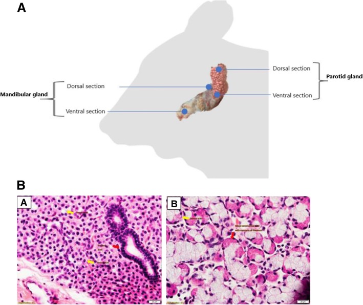
Fig. 2.
a Salivary glands sampling. After euthanasia, the head was positioned upside down and the skin between jaws was incised using sterile disposable scalpel. Then, diagonal incision was made from the ear to join the first incision and the skin was removed from one side to expose the adjacent tissues. Fatty tissue was incised at the site of targeted salivary glands. Parotid and mandibular glands were located at one side and two samples were collected at dorsal and ventral anatomical sections from each gland. b: a: Parotid gland; Pure serous acini consisting of rectangular granular cells with central nuclei. Central lumen hardly visible (yellow arrow). Striated duct with columnar cells with central nuclei and basal-striated appearance (red arrow). b Mandibular gland; Pure serous acini consisting of triangular granular cells with their base directed outwards and basal nuclei (yellow arrow). Mixed seromucous acini with crescents of Giannuzzi (red arrow). Bar length 20 um

