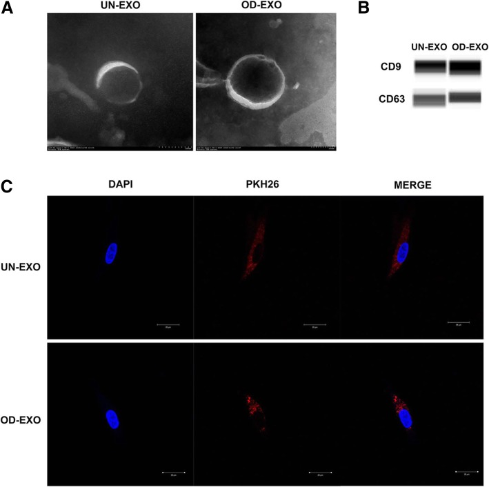
Fig. 2.
Endocytosis of UN-Exo and OD-Exo by DPSCs. a The morphology of UN-Exo and OD-Exo was determined by transmission electron microscopy. b Automated western blot analysis revealed that exosomal markers CD9 and CD63 were expressed in the UN-Exo and OD-Exo. c Endocytosis of exosomes by DPSCs was visualized by fluorescent labeling with PKH26

