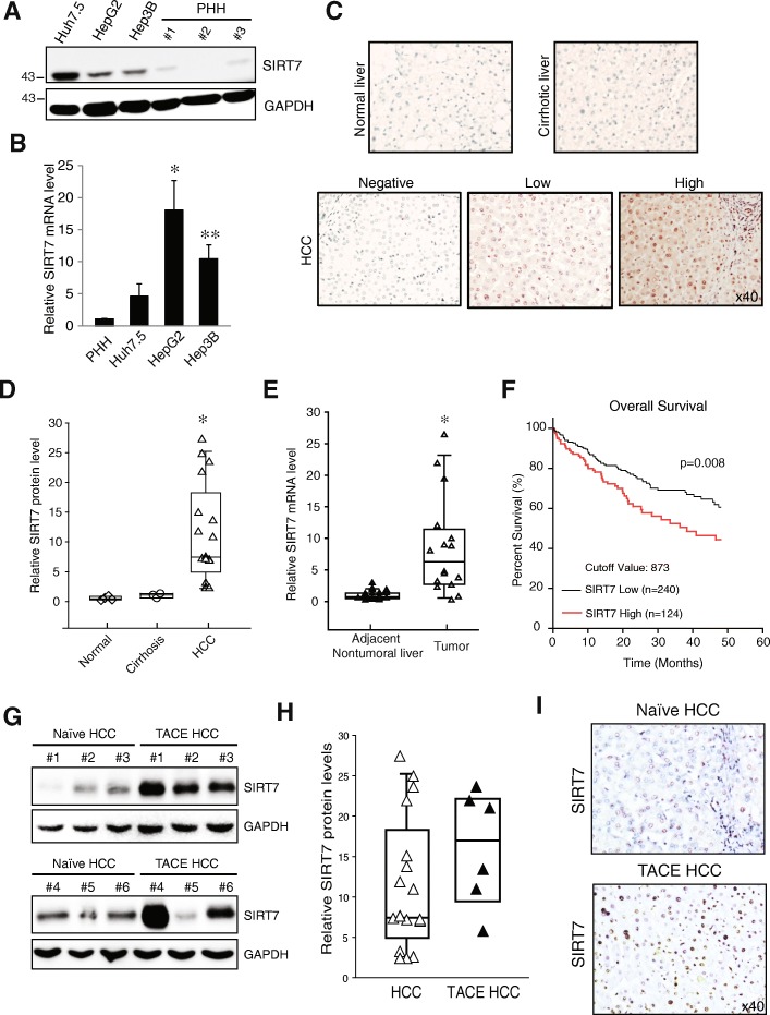
Fig. 1.
SIRT7 expression in human HCC. Protein (a) and mRNA (b) levels of SIRT7 from isolated primary human hepatocyte (PHH) and HCC cell lines. c Representative IHC staining of SIRT7 in normal, cirrhotic and HCC liver sections. d Quantitative analysis of SIRT7 protein levels in normal (n = 4), cirrhotic (n = 3) and HCC (n = 17). The data is presented as fold increase compared with normal liver and was normalized to GAPDH. *P < 0.05 vs normal, Student’s t-test. e RT-PCR analysis of SIRT7 mRNA levels in 20 paired nontumoral and HCC tissues. *P < 0.05, Student’s t-test. f Kaplan–Meier analysis of overall survival in 364 liver cancer patients based on SIRT7 expression. g and h Western blot analysis of SIRT7 protein levels (g) and quantitative analysis of SIRT7 protein levels in HCC as in D (n = 17) and TACE treated HCC (n = 6) (h). The data is presented as fold increase compared with normal liver and was normalized to GAPDH. i IHC staining of SIRT7 in Naïve and TACE treated HCC tissues

