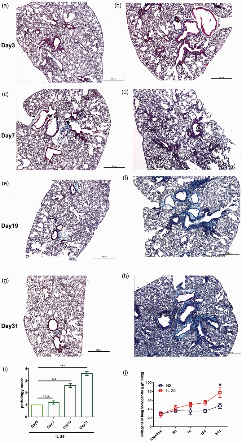
Figure 3.
Masson and soluble collagen assay show increased collagen deposition following mIL-25 intranasal instillation. Mice were intranasal injected with of mIL-25 or saline and were sacrificed at days 3, 7, 19, and 31 for lung tissue and homogenate collection. Representative low-power histology images show extensive collagen deposition by using Masson’s trichrome stain at indicated time points (a to h). Lung pathology score is shown in panel (i), data are means ± SEMs of five mice at indicated time points, NS: no significance, *P < .001 at days 7, 19, and 31 compared with day 3. Total lung soluble collagen in lung homogenate is determined by Sircol assay kit (j). Data are shown as means ± SEMs (N = 5 per group per experiment) *P < .05 compared between saline and IL-25 group at indicated time points, as calculated by ANOVA with Bonferroni post hoc test. IL-25: interleukin 25. (A color version of this figure is available in the online journal.)

