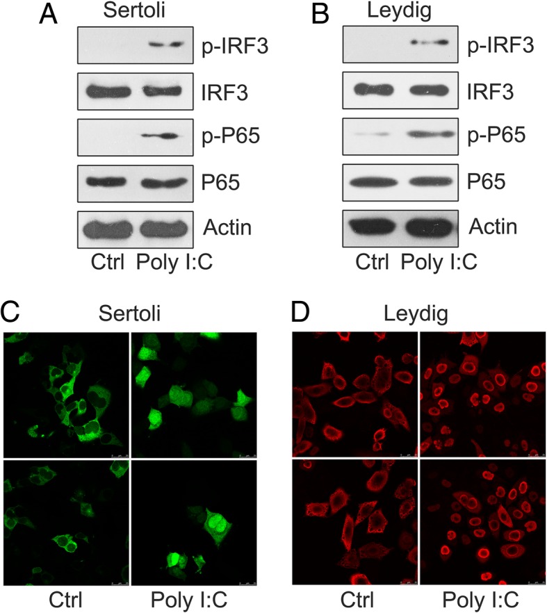
Fig. 2.

Poly I:C triggered NF-κB and IRF3 expression. a Sertoli and b Leydig cells were treated for 6 h with 2 μg/mL of poly I:C. Lysates were subjected to western blot analysis and probed with antibodies for phosphorylated IRF3 (p-IRF3) p-P65, total P65, and IRF3. β-Actin served as a loading control. c and d) IFA was used to study the nuclear translocation of IRF3 and P65. Leydig and Sertoli cells were treated with 2 μg/mL of poly I:C. Distribution of P65 and IRF3 inside the cells was assessed with indirect IFA. Images were obtained for P65 and IRF3 translocation in Leydig (red) and Sertoli (green) cells after 8 h treatment. Cells with no transfection served as controls (Ctrl). Images were independently obtained for at least three experiments
