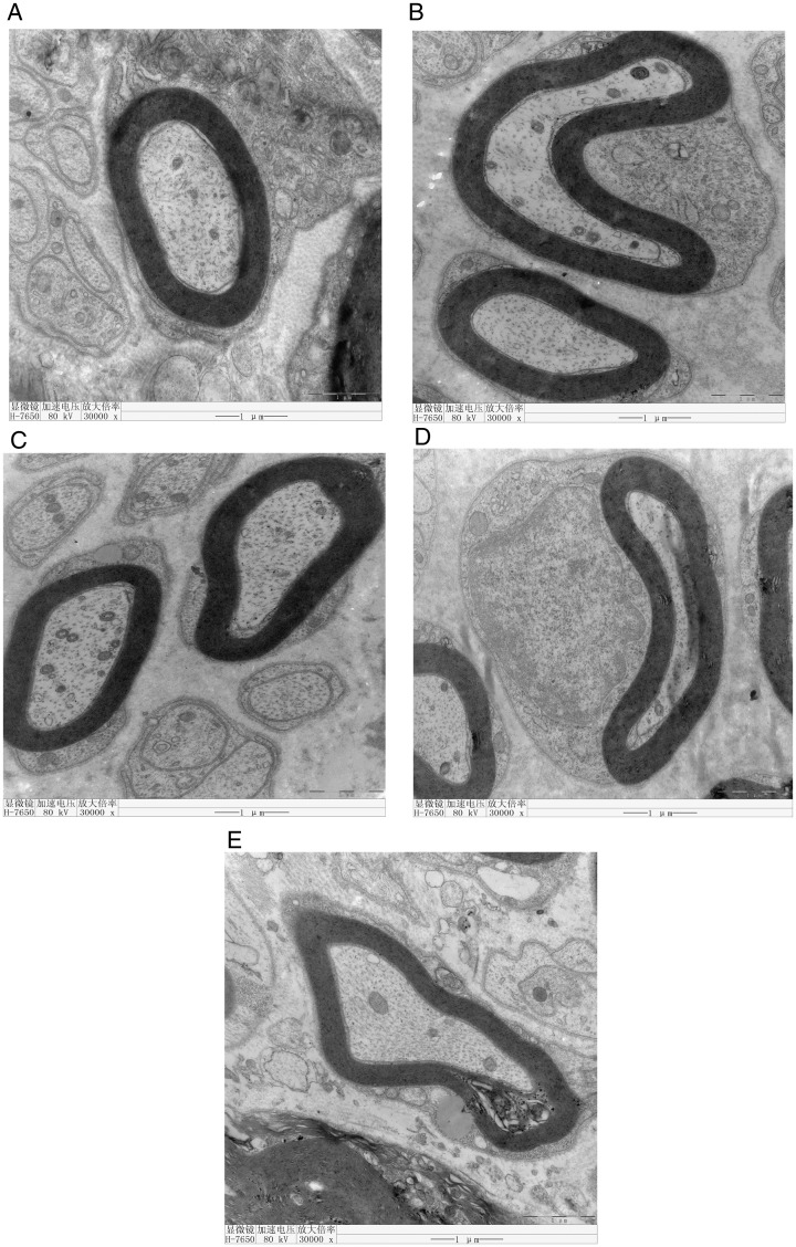
Figure 1.
Electron microscopic examination of Groups A, B, C, and D shows that the myelin lamella of nerve fibers is rounded or oval with a compact structure. The structure of Schwann cells is clear and the cytoplasm is uniform. The ultrastructure of mitochondria and microfilaments is clear. However, the myelin lamella is loose and vacuolated in Group E.

