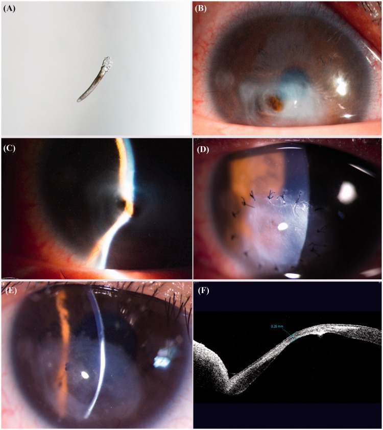
Figure 1.
Patient 1. (a) Microscopic picture showing Demodex in the epilated eyelashes. (b) Preoperative slit-lamp appearance of the right eye showing conjunctival congestion, corneal thinning, and corneal perforation in the inferotemporal region with iris prolapse as well as (c) a shallow anterior chamber. (d) Postoperative image showing the trimmed lenticule sutured with interrupted 10-0 nylon sutures and (e) the intact graft with no sign of graft rejection after suture removal. (f) Anterior-chamber optical coherence tomography showing a corneal thickness of 0.28 mm at the site of the perforation after the surgery.

