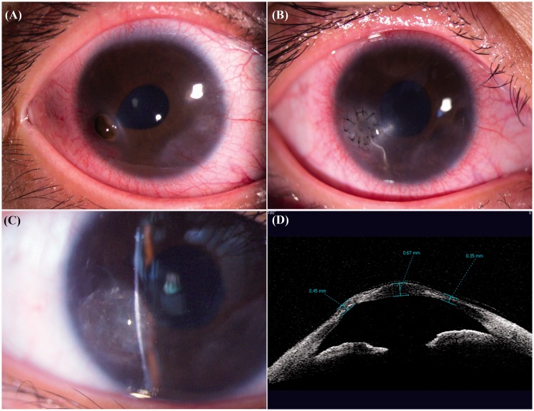
Figure 2.
Patient 2. (a) Preoperative slit-lamp appearance of the right eye showing ciliary injection, an irregular pupil, and perforated cornea in the inferior part of the temporal region with prolapse of the iris. (b) Postoperative image showing an intact suture with the graft in position. (c) Image after removal of the suture with a sealed corneal perforation and no sign of rejection. (d) Anterior-chamber optical coherence tomography showing a corneal thickness of 0.45 mm at the site of the perforation after the surgery.

