Figure 6.
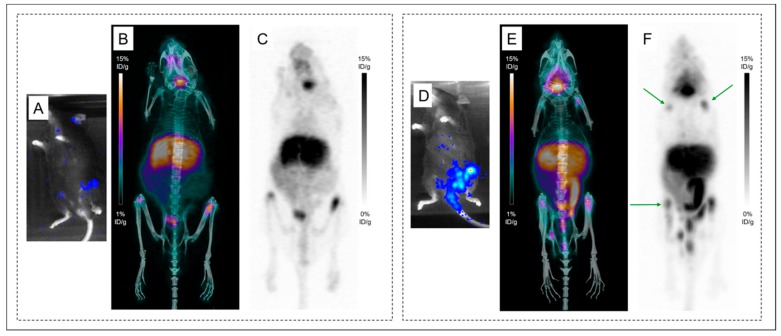
Altered contrast in the disseminated model imaged with 89Zr-DFO-9E7.4 at 24 h post-injection. Bioluminescence imaging (A) revealing lesions in both femurs. Maximum intensity projections of PET and CT imaging with 89Zr-DFO-9E7.4 at 24 h post-injection (PI) (B) and maximum intensity projections of PET imaging with 89Zr-DFO-9E7.4 at 24 h PI (C) showing uptake in both femurs (Mouse 16). Bioluminescence imaging (D) revealing lesions in the sacrum, left femur and both iliac wings. Maximum intensity projections of PET and CT imaging with 89Zr-DFO-9E7.4 at 24 h PI (E) and maximum intensity projections of PET imaging with 89Zr-DFO-9E7.4 at 24 h PI (F) showing uptake in the sacrum, left femur and both iliac wings (Mouse 17). Multiple false-positive osseous foci were also observed (indicated by green arrows).

