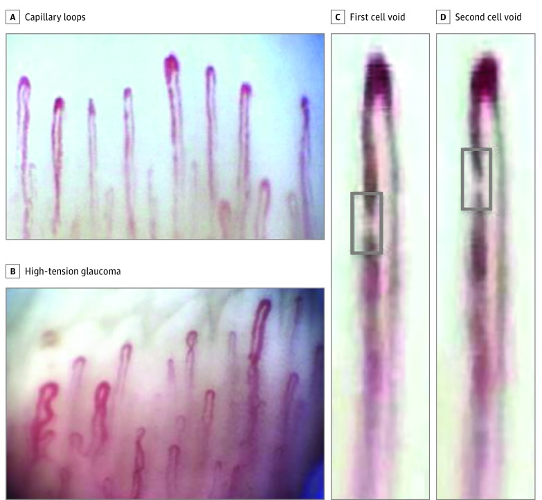
Figure 1. Video Nailfold Capillaroscopy Images Showing Capillary Loops (Inverted U-Shape).
A, Control participant. B, Patient with high-tension glaucoma. C and D, Magnified capillary showing plasma gap/blood cell void (gray box and seen as the white band) movement over 2 consecutive image sequence frames in a control participant. Flow was 64.56 picoliters per second.

