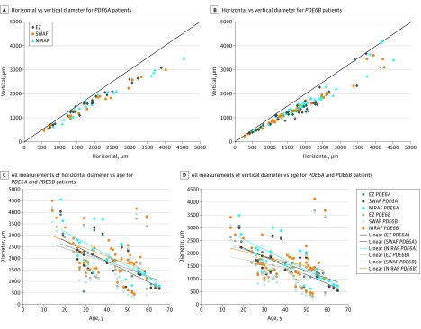
Figure 3. Longitudinal Structural Changes of Spectral-Domain Optical Coherence Tomography (SD-OCT), Short-Wavelength Fundus Autofluorescence (SWAF), and Near-Infrared Fundus Autofluorescence (NIRAF) Images of Patients With PDE6A and PDE6B Mutations.
Horizontal and vertical diameters measured for ellipsoid zone (EZ), inner SWAF, and outer hyperautofluorescent parafoveal rings in patients with PDE6A (A) and PDE6B (B) mutations showing that the vertical diameter is smaller than the horizontal diameter and becoming equal with degeneration progression. All SD-OCT, SWARF, and NIRAF horizontal (C) and vertical (D) measurements of both groups plotted on the same chart show measurements within comparable ranges and clustering into parallel trend lines.

