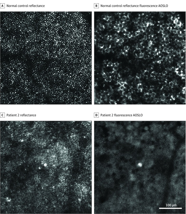
Figure 2. Adaptive Optics Scanning Light Ophthalmoscopy (AOSLO) Imaging Near the Fovea (0.5 mm Eccentricity).
A and C, Confocal reflectance AOSLO. B and D, Fluorescence AOSLO. Patient 2 shows no detectable photoreceptor or retinal pigment epithelial cells (C and D) compared with a matched location in a normal eye (A and B). The location marked by blue box 1 in Figure 1 corresponds to the AOSLO imaging location here in C and D.

