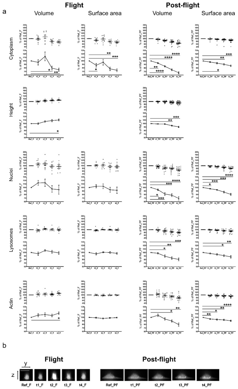
Figure 3.
Microgravity-induced changes of cellular and sub-cellular structures. (a) Averaged values and statistical evaluation of the single cell analyses. Living human primary macrophages were stained with Nuclear Violet (nuclei staining), Calcein (cytoplasm staining), LysoBrite (lysosome staining), and SiR-actin (F-actin staining) and exposed to microgravity during the TEXUS-54 suborbital ballistic flight. 10 min before the flight and at four times during the flight, confocal microscopic pictures were taken with the confocal laser spinning disk fluorescence microscope FLUMIAS. Additionally, post-flight ground controls were performed. Volume, surface area, and the height of single cells (upper part of each graph) were quantified (software based) after correction of the laser-induced bleaching effect. Averaged values are displayed in the lower parts of the graphs. Error bars represent SEM. p-values ≤ 0.05 were considered as significant (p ≤ 0.05 = *, ≤0.01 = **, ≤0.001 = ***, ≤0.0001 = ****) (b) Sagittal z-stack images of representative example cells stained with the cytoplasmic marker Calcein for all time points of flight and post-flight acquisitions. Scale bar: 20 µm.

