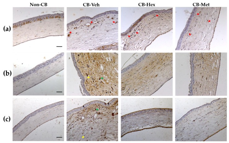
Figure 2.
Micrograph of Met and Hex extracts of C. argyrosperma seed in the CB compared to the non-CB and CB-Veh groups. (a) Nuclei stained with anti-NF-κB p65 (red arrows). (b) Staining intensity for interleukin-1β (IL-1β) along with the corneal thickness. (c) Cyclooxigenase-2 (COX-2) staining in the studied groups. Arrowheads indicate the cytoplasmic distribution of NF-κB p65. Yellow and green arrows represent the minimum and maximum staining intensity, respectively, considered for software analysis. (Scale bar = 100 μm). CB: chemically burned corneas; CB-Veh: CB corneas treated with the vehicle; CB-Hex: CB corneas treated with hexanic extract; CB-Met: CB corneas treated with methanolic extract.

