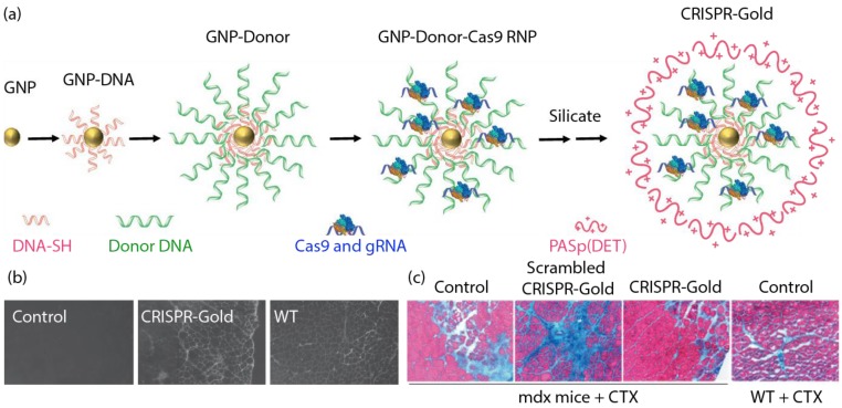
Figure 5.
(a) Generation of CRISPR-Gold. (b) CRISPR-Gold-injected muscle of mdx mice showed dystrophin expression (immunofluorescence), whereas control mdx mice did not express dystrophin protein. (c) CRISPR-Gold reduces muscle fibrosis in mdx mice. Trichrome staining was performed on the tibialis anterior muscle cryo-sectioned to 10 μm two weeks after an injection of CRISPR-Gold. CTX was co-injected in all three groups of mdx mice. Images were acquired at the areas of muscle injury and regeneration. Fibrotic tissue appears blue, while muscle fibers appear red. Wild-type mice treated with CTX were analyzed five days after injection. (Adapted with permission from Ref. 82 Copyright (2017) Macmillan Publishers).

