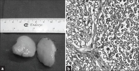
Figure 2.

(a) Cut section of the tumor, 2 cm × 2.8 cm, fleshy and smooth in appearance. (b) Highly cellular tumor, lobules separated by fibrocollagenous septae, individual cell shows scanty cytoplasm, uniform round-to-oval vesicular nuclei with high mitotic activity (H and E, ×40)
