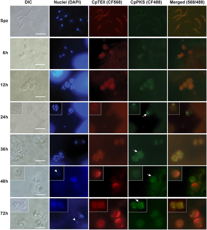
Figure 4.
Indirect immunofluorescence microscopic detection of CpTEII and CpPKS protein in different life stages of C. parvum. Anti-CpTEII antibodies stain highly on some later stage parasites (48 and 72 h post-infection), which are probably macrogamonts with large or unstainable nuclei by DAPI (arrowhead), and relative weak signal can be observed on sporozoites and other parasites. CpPKS staining signal is very faint on sporozoites and weak in 6 and 12 h postinfection stage parasites, and become stronger on some 24, 36, 48, and 72 h postinfection stage parasites (arrow). DIC, differential interference microscopy; DAPI, 4′,6-diamidino-2-phenylindole for staining nuclei; Merged, superimposed images of CpTEII and CpPKS. Bar = 5 μm.

