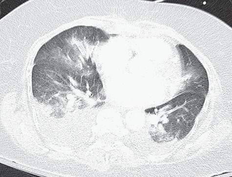
Figure 3.

CT angiography of the chest with intravenous contrast showing no filling defects within the pulmonary vasculature, and re-demonstration of multifocal ill-defined patchy/fluffy airspace consolidative and ground glass opacities within the bilateral lungs, favored to represent pulmonary edema.
