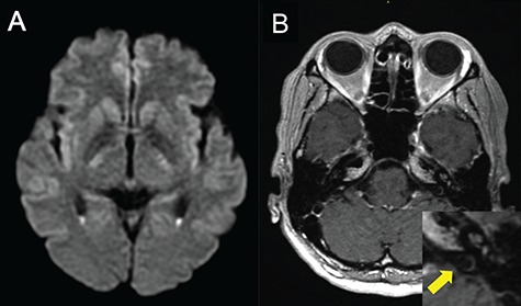
Figure 3.

MRI shows PV (a) and suppurative labyrinthitis (b). (a) Diffusion-weighted image reveals hyper-intense lesions with a fluid–fluid level in the trigones of the bilateral lateral ventricles; (b) gadolinium-enhanced T1-weighted image reveals increased signals in the cochlea of the left inner ear lesions (yellow arrow of a zoomed-in image).
