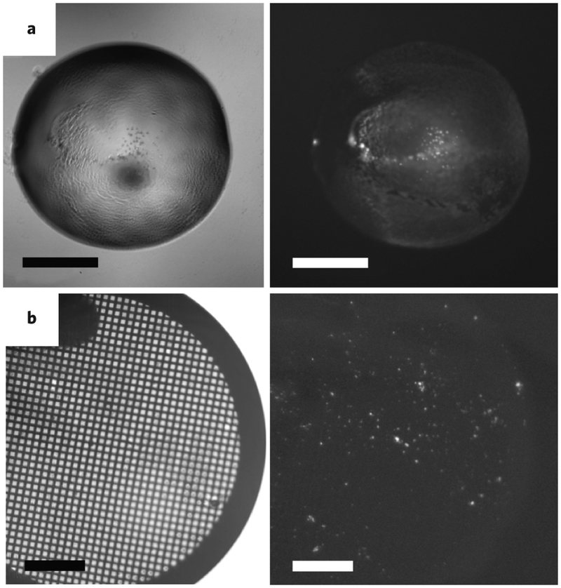
Figure 5: Crystal identification and sample preparation for MicroED.

(A) Frequently, identification of microcrystals in drops that appear to have cloudy precipitates is difficult by visible light (left panel); however, when the drops are imaged using UV fluorescence, the presence of microcrystals is clearly seen by sharp glowing spots (right panel). (B) UV fluorescence can also be used to visualize the presence of microcrystals deposited on the EM grid during sample preparation for EM. Scale bars represent 500 μm.
