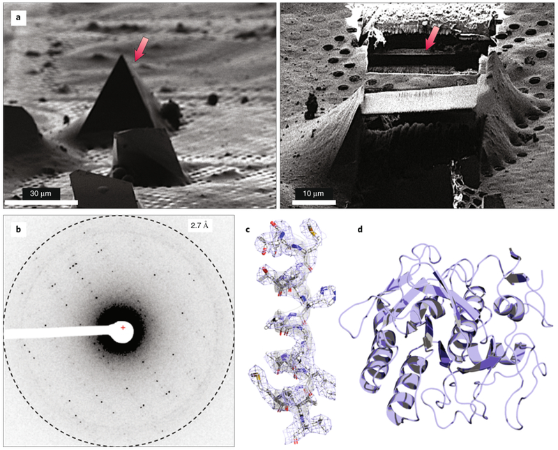
Figure 6. Cryo-FIB milling of a thick crystal.

(A) Sample preparation for MicroED using a cryo-FIB to mill down thick crystals to a few hundred nanometers (left and right). (B) Following cryo-FIB milling, the grid would be loaded into the TEM and diffraction data would be collected from the thin lamella. (C-D) Final structure of proteinase K.
