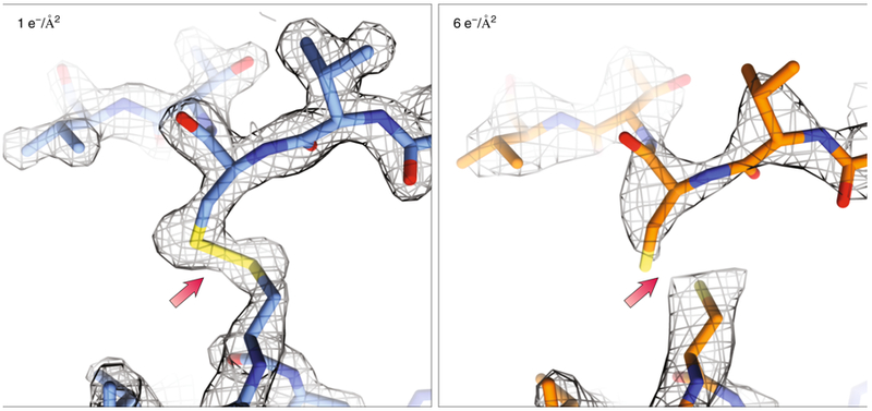
Figure 8: Dynamics probed in response to radiation damage.

When less than 1 e−/Å2 (left) was used for structure determination of Proteinase K (1.7Å, PDB ID: 6CL7), local radiation damage was minimal. When higher doses (right) were used (2.8Å, PDB ID: 6CLA) the damaging effects of the beam can already be seen with the breakage of disulfide bonds and the lower attainable resolution. Density maps (2Fo-Fc) in gray are contoured at 1.5σ.
