
Fig. 2. Organelle division by ER MCSs.
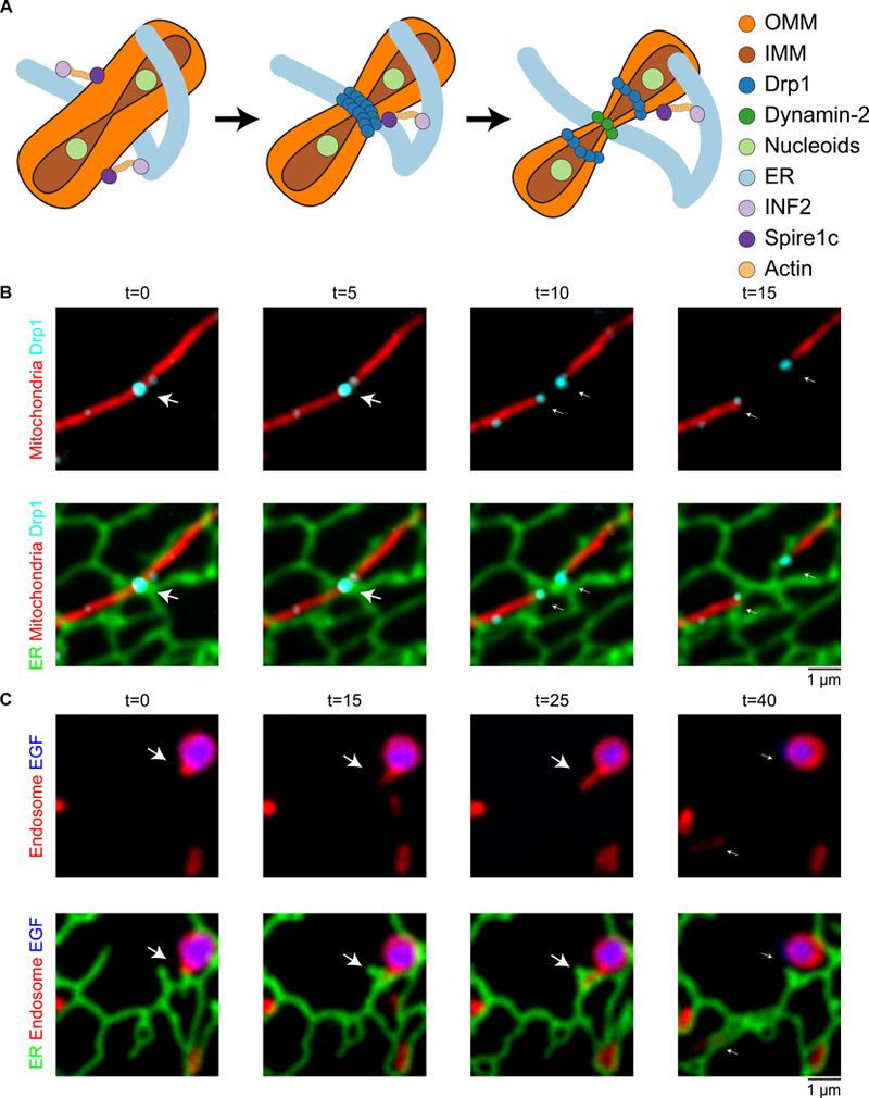
(A) Model of factors involved in ER-associated mitochondrial constriction and division in animal cells: ER MCSs and actively replicating nucleoids define where 1) inner mitochondrial membrane constriction followed by 2) outer mitochondrial membrane (OMM) constriction. OMM constriction requires the activities of INF2, Spirelc, and polymerized actin, followed by the sequential activity of Drp1 and Dnm2 to drive OMM constriction and division. (B) ER tubules define the position of mitochondrial constriction and division. Time-lapse live-cell imaging of the ER (green) and mitochondria (red) in a COS-7 cell shows the division machinery Drp1 (cyan) is localized to the position where an ER tubule crosses over a mitochondrial constriction (panel 1 and 2). As the mitochondria divides, the Drp1 punctum splits and the ER tubules bridge the gap to maintain contact with both Drp1-labeled ends on daughter mitochondria (arrow). Images provided by J. Lee. (C) Dynamic ER tubules are recruited and rearrange around endosome cargo sorting domains to promote endosome fission. Time-lapse live-cell imaging of the ER (green), late endosomes (red) and EGF cargo (blue) shows an endosome bud growing through an ER ring (arrow) and as the ring closes, the bud undergoes fission (compare 3rd and 4th time frame). Image reproduced from (61).