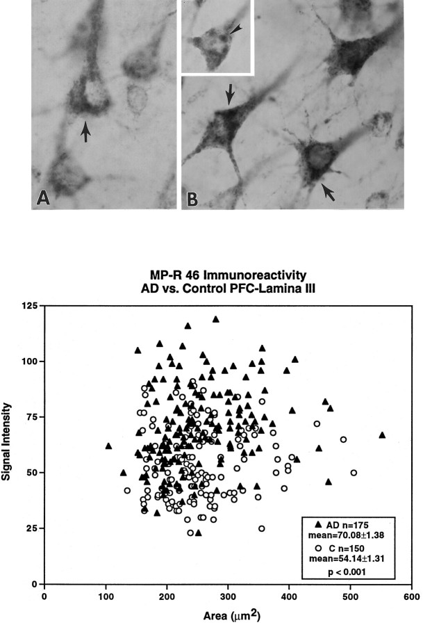
Fig. 5.
Levels of MP-R46 are elevated in neurons of AD brains. Immunocytochemical studies using antibody directed against MP-R46 show increased levels of this receptor (arrows) in lamina III neurons in the AD brains (B) than in controls (A). (The cells in Arange in optical density from 57 to 67, control mean = 54.14 ± 1.31; those in B range in optical density from 71 to 86, AD mean = 70.80 ± 1.38; p < 0.001. Percentage difference above the mean optical density was equivalent for neurons inA vs B.) In AD brains displaying lower levels of immunostaining, MP-R46 immunolabeling could be visualized clearly within large vacuolar profiles (B, inset, arrowhead) that resembled the abnormally large rab5-positive early endosomal profiles. Semiquantitative morphometric analysis from sections of the prefrontal cortex revealed that cortical pyramids in laminae III of the AD brains ( filled triangles) exhibit increased MP-R46 densities per cross-sectional area as compared with age-matched controls (open circles). Magnification inA, B, B inset, 3600×.

