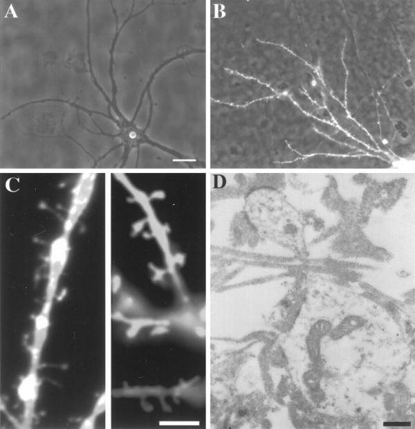
Fig. 6.
Development of dendritic spines in the presence of innervation. Micrograph of hippocampal neurons growing off (A) and on (B) the entorhinal axonal net. Only neurons growing in contact with the entorhinal axons develop numerous spines. C, Fluorescent micrograph showing spines of DiI-stained neurons at higher magnification. D, Electron micrograph of a spine. Scale bars: A, B, 25 μm; C, 5 μm;D, 0.5 μm.

