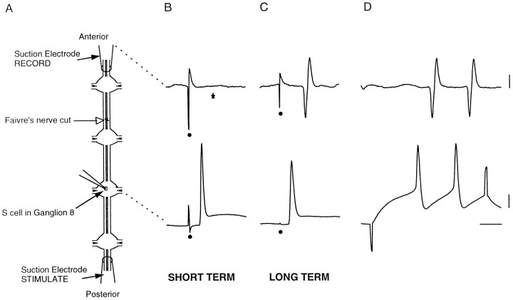
Fig. 3.
Extracellular stimulation and recording demonstrate disconnection and reconnection of S-cells.A, Diagram of preparation for recording, depicting locations of suction electrodes at the ends of the connectives, an intracellular microelectrode in S8, and the lesion site in the medial connective (open arrow) anterior to ganglion 7. B, For short-term specimens, 5 d after surgery, the impulses elicited in the S-cell by stimulating the connectives extracellularly with the posterior suction electrode did not propagate across the lesion to the anterior suction electrode. Stimulus artifact is marked (•), and the usual location of the propagated extracellular action potential, missing here, is marked (★). C, For long-term specimens, here 78 d, the S-cell impulses propagated across the lesion. In D, depolarization of S8 produced a pair of impulses that were also recorded extracellularly. The recording sites on the diagram inA are linked by dashed lines to the corresponding voltage traces in B–D. Calibration bars at right for B and C, 1 mV, extracellular trace; 10 mV, intracellular trace; and 10 msec.

