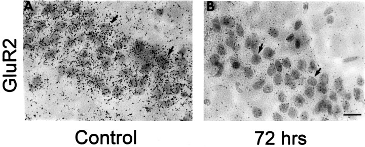
Fig. 5.
Changes in GluR2 mRNA expression are cell-specific. Emulsion-dipped coronal sections of the hippocampus from control and experimental animals 72 hr after ischemia show silver grains densely clustered over CA1 pyramidal neurons. A, Sections of control brain revealed dense clusters of silver grains overlying individual pyramidal neurons in the CA1; virtually all neurons in the field exhibited intense labeling. B, Seventy-two hours after ischemia, GluR2 labeling was dramatically reduced for all CA1 neurons. Sections were counterstained with hematoxylin and eosin. Arrows indicate representative pyramidal neurons. At this time, histological analysis showed no cell loss (see Fig. 1).

