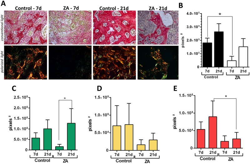
Fig 3. Birefringence analysis of collagen fibers in the alveolar sockets post tooth extraction (E sides) in control and ZA treated mice.
Senescent 129 Sv-WT female mice received IP injections of 0.9% saline solution (Vehicle) or 250 μg/Kg once a week and upper right incisor were removed after 4 weeks of Vehicle or ZA treatments. Mice were euthanized for maxillary bones removal after 7days and 21days post tooth extraction. A)Representative transversal sections alveolar socket upon polarized and conventional light, to evaluate collagen fibers maturation in the different experimental groups and periods. As visualized upon polarized light, green birefringence color indicates thin fibers; yellow and red colors at birefringence analysis indicate thick collagen fibers. Original magnification was 40x. Intensity of birefringence measured from Image-analysis software (AxioVision, v. 4.8, CarlZeiss) to identify and quantify total area of collagen fibers (B) and area of collagen from each birefringence color (pixels2) (C-green, D-yellow and E-red). Results are presented as mean and SD of pixels2 for each color in the birefringence analysis. Symbol * indicates a statistically significant difference vs. control (p<0.05).

