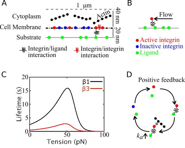
Fig 2. Schematic illustration of the model system.

(A) Side view of the computational domain, where single-point, two-state integrin particles diffuse and assemble laterally. Upon activation (from blue to red), integrins can establish interactions (black spring) with ligands (green particles) and other active integrins (red springs). (B) Schematics of the system with actin flow: a force mimicking actin flow is exerted on ligand-bound integrins, parallel to the substrate, and builds tension on the integrin/ligand bond. (C) Lifetime versus tension curves of catch bonds used to mimic β-1 (black) and β-3 (red) integrins. (D) Schematic illustration of the positive feedback between actin filament binding and integrin activation. Once an integrin particle is bound to a ligand, it can establish interactions with actin. Upon binding actin, its activation rate increases. Upon deactivating and unbinding, its propensity to become active again increases, leading to increased probability of binding new ligands and actin filaments. This results in a positive feedback between ligand binding and engaging the actin cytoskeleton.
