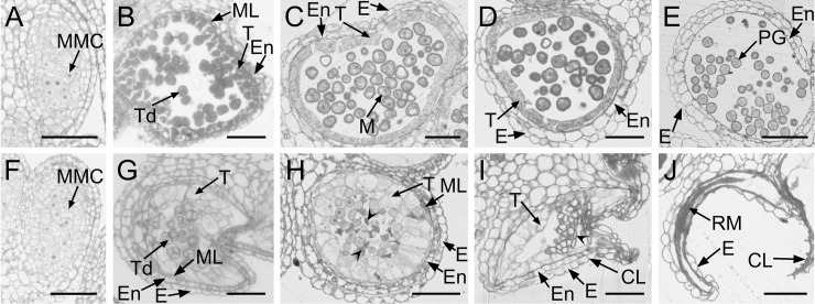
Fig 3. Anther and microspore development in the Ogura-CMS line and its maintainer fertile (MF) line of turnip.
(A-E) Semi-thin sections of the MF anthers. (F-J) Semi-thin sections of the Ogura-CMS anthers. (A, F) Microspore mother cell stage. (B, G) Tetrad stage. The young microspores are surrounded by a callose wall, a tapetum, a middle layer, an endothecium, and an epidermis from the inside out at the tetrad stage. The tapetum in (G) swells at the center of the locule. (C, H) Uninucleate microspore stage. The middle layer persisted in (J). The aborted microspores indicated by arrowheads in (J) was surrounded by a swollen tapetal layer. (D, I) Bicellular stage. The collapse of anther locule is obvious with the aborted microspores indicated by arrowhead in (I). (E, J) Dehiscent stage. Endothecium layer is absent in the surrounding walls and remnants of the aborted microspores adhere to the inner face of the epidermis in (J). CL, collapsed locule; E, epidermis; En, endothecium; M, microspore; ML, middle layer; MMC, microspore mother cell; PG, pollen grain; RM, remnants of microspores; T, tapetum; Td, tetrads. Bars = 50 μm.

