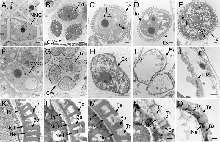
Fig 4. Transmission electron microscopy observation of microspore development in the Ogura-CMS line and its maintainer fertile (MF) line of turnip.
(A-E) Images of microspore development in the MF line from the microspore mother cell stage to the mature pollen stage. (F-J) Images of microspore development in the Ogura-CMS line from the microspore mother cell stage to the mature pollen stage. (A, F) Microspore mother cell stage. (B, G) Tetrad stage, showing four young microspores surrounded by the callose wall. (C, H) Uninucleate microspore stage, showing the intine and the germinal apertures commenced in (C). (D, I) Bicellular stage, showing the degenerated microspores in (I). (E, J) Mature pollen stage, showing the mature pollen grain in the MF line (E) and the remnants of microspores in the Ogura-CMS line (J). (K-M) Magnified images of pollen wall in (C-E), showing the multilayered structure. (N, O) Magnified images of pollen wall in (H, I), showing the incomplete-developed exine layer and the absence of inine layer. Ba, baculum; CW, callose wall; Ex, exine; GA, germinal aperture; In, intine; M, microspore; MMC, microspore mother cell; Ne I, nexine I; Ne II, nexine II; PG, pollen grain; RM, remnants of microspores; Td, tetrads; Te, tectum; Tr, tryphine. Bars = 2 μm in (A-J), 0.2 μm in (K-O).

