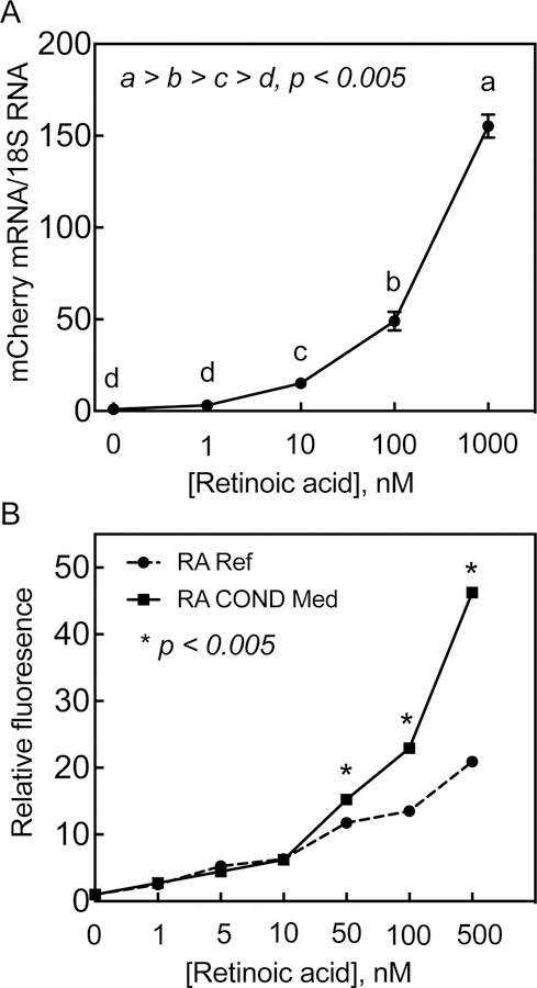
Figure 5. Red fluorescent protein (RFP) is expressed quantitatively in HEK293T 1A1 cells in response to RA.
A. HEK293T 1A1 cells grown in 12-well plates were incubated with RA at different concentrations for 24 h after which the cells were washed and collected for total RNA isolation. The total RNA samples were then subjected to RT-PCR analysis for expression of mCherry RNA in triplicate using 18S ribosomal RNA expression as the internal control. B. HEK293T 1A1 cells were grown in 12-well plates then incubated 24 h with either RA at different concentrations or 24-h RA treated conditioned media from HepG2 cells. For conditioned media preparation, HepG2 cells were incubated RA at different concentrations in EMEM full growth media for 24 h. The media were collected and then incubated at 50% with HEK293T 1A1 cells without addition of any RA. For reference standard, the HEK293T 1A1 cells were incubated with 50% HepG2 cell 24-h vehicle-treated conditioned media with added RA at different concentrations for 24 h. The HEK293T 1A1 cells in duplicate wells were then trypsinized and collected for flow cytometry for the cells expressing RFP.

