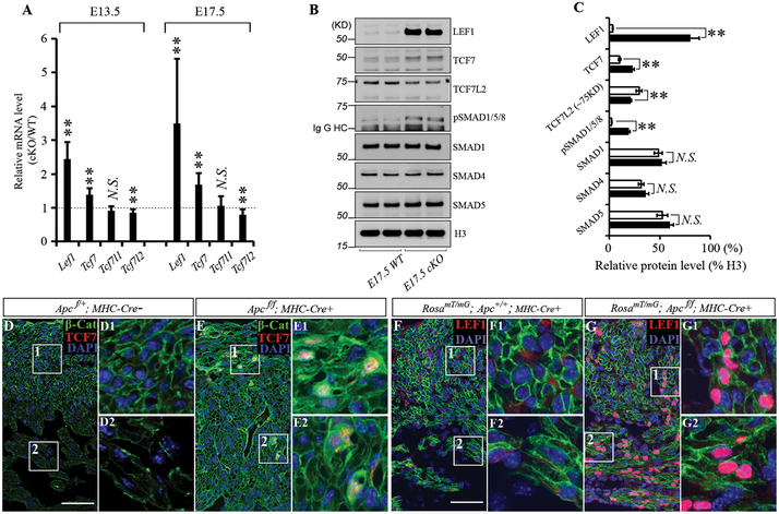
Figure 3. Alteration of β-catenin partners by Apc deletion.
A, A bar graph shows the ratio of Tcf/Lef mRNA expression levels of hearts between Apc knockout (KO) animal and their wild type (WT) siblings at E13.5 and E17.5 by q-PCR. B, Upregulation Lef1 and Tcf7, but downregulation of long form of Tcf7l2 (~75 kDa) upon cardiac Apc deletion at E17.5 by western blot. Short form of Tcf7l2 (~54 kDa) is not changed. pSmad1/5/8 is also increased by ablation of Apc. Uncropped images of Western blots are displayed in Fig. S6. C, Band intensities are quantified by ImageJ and normalized to Histone 3. D and E, Tcf7 was not detected in WT (D), but was induced in Apc KO mice and colocalized with nuclear β-Cat in CMs (E) at E17.5. F and G, Tomato hetero hearts revealed efficient conversion of membranous red (not shown well due to weaker signal than that of Lef1) to green fluorescence by αMHC-Cre. Lef1 was not detected in WT (F), but was dramatically elevated in Apc KO mice (G) at E17.5. Scale bars=50μm. Data represent mean ± SD. N=4 independent experiments.

