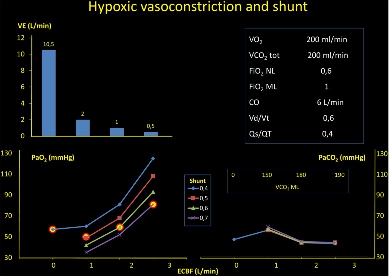
Fig. 1.
In the right square, we present the starting conditions of this analysis. In the left upper panel, we present the decrease of the total ventilation to maintain an unchanged PCO2 (lower right panel) when the extracorporeal blood flow is increased. In the left lower panel, we show which would be the arterial PO2 as a function of the extracorporeal blood flow, if the shunt fraction would be unmodified. As shown, an extracorporeal blood flow of 1.5 L/min, if the shunt increases to 0.4 to 0.7 (a common finding in this condition), the PaO2 increase, if any, is negligible

