
Fig. 7.
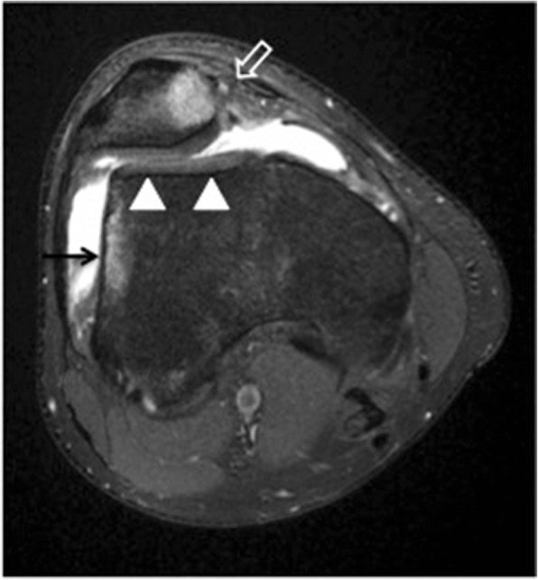
Transient lateral patellar dislocation. Axial PDFS MR image showing MPFL disruption (open arrow) and trochlear dysplasia (arrowheads). There is edema of the medial patella and of the lateral femoral condyle (arrow), consistent with bone contusion due to recent lateral patellar dislocation