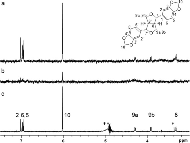
Figure 2.

STD-NMR analyses of sesamin and GST-fused Steroleosin B. ¹H STD-NMR spectra of sesamin with GST-fused Steroleosin B (a) or GST (b) at a ligand to protein molar ratio of 60:1; (c) one-dimensional ¹H NMR spectra of sesamin. Signals were assigned to the sesamin structure. Those of C2′, C5′, C6′, C8′, C9′ and C10′ overlapped exactly with those of C2, C5, C6, C8, C9 and C10, respectively, due to the C2 symmetric structure of sesamin; *impurity; **C7- and C7′-protons in this area disappeared due to the suppression of non-deuterated water protons with similar chemical shifts.
