Figure 4.
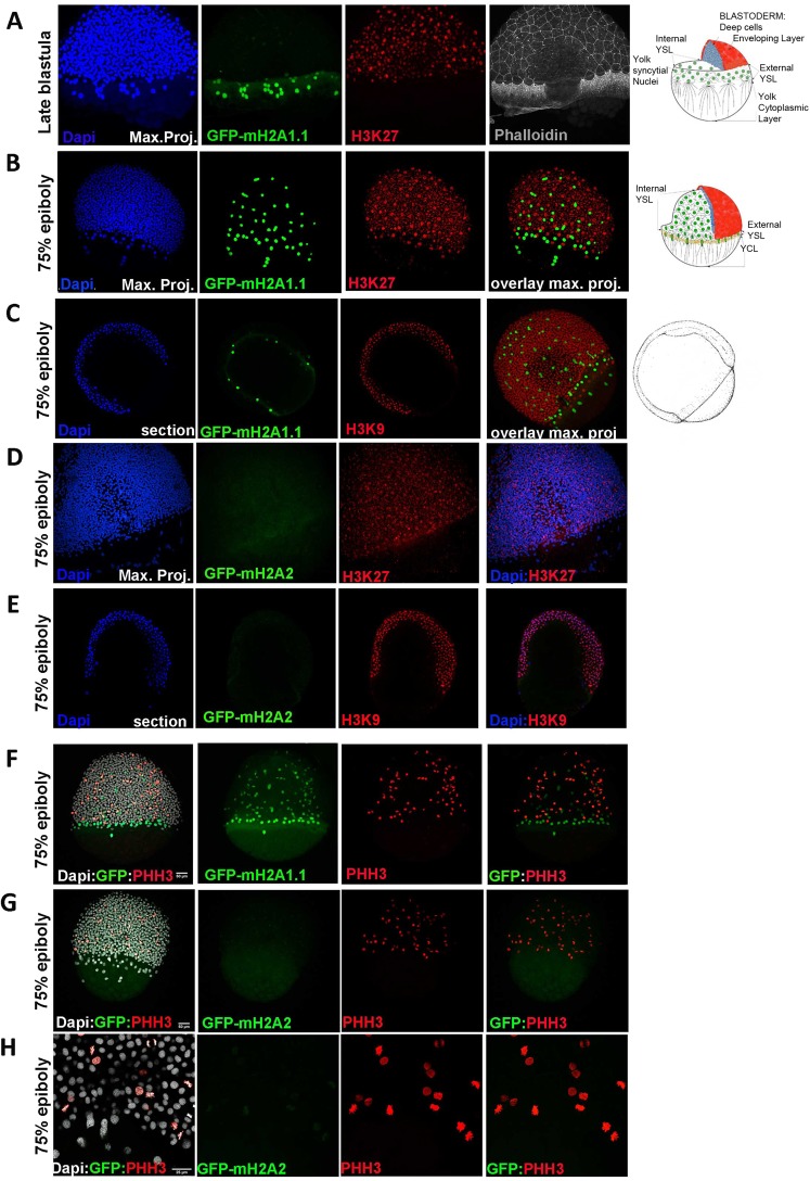
mH2A isoforms differential colocalization with heterochromatin and mitotic marks in zebrafish embryos at epiboly stage. Transgenic zf embryos expressing mH2A1:GFP-mH2A1.1 (A–C,F) or mH2A2:GFP-mH2A2 (D,E,G,H) at late blastula (A) or 75% epiboly (B–H) stages were analysed using immunohistochemistry to detect the heterochromatin markers trimethyl Histone3 lysine K27 and K9 (H3K27me3 and H3K9me3) (A–E) or mitotic nuclei phosphohistone H3 (pHH3) (F–H). DAPI and Phalloidin-conjugate were used for nucleus and cytoskeleton labelling respectively. (A–C) Confocal microscope image projection of late blastula (A) and 75% epiboly stage (B) transgenic mH2A1:GFP-mH2A1.1 embryo shows YSL localization of GFP positive cells and lack of colocalization with H3K27me3. Apical pole and vegetal pole are pointed in the image. To the right of (A–C) there is a schematic illustration of the organization of the cortical cytoplasm of the yolk cell in relation to other cell types in zebrafish embryo during epiboly (draws at late blastula and 60% epiboly respectively). (C) Confocal microscope image section also shows absence H3K9m3 signal in the nuclei of YSL cells, where GFP-mH2A1.1 is expressed. (D,E) Confocal microscope image projection (D) and section (E) of 75% epiboly stage transgenic mH2A2:GFP-mH2A2 embryo stained with heterochromatin markers H3K27me3 and H3K9me3 shows heterochromatin labelling in the whole embryo while GFP (mH2A2) expression is still very weak. (F–H). Confocal microscope image projection of 75% epiboly stage transgenic mH2A1:GFP-mH2A1.1 (F) or mH2A2:GFP-mH2A2 (G,H) with labelled pHH3 mitotic nuclei. Figure show non-mitotic mH2A1 positive YSL nucleus and proliferative cells within the embryo body. (H) Higher magnification of images of mH2A2:GFP-mH2A2 transgenic fish clearly showing pHH3 positive cells while there is a low level GFP-mH2A2 expression (H). Each labelling assay was conducted in triplicate with n = 40–50 embryos/immunolabeling.

