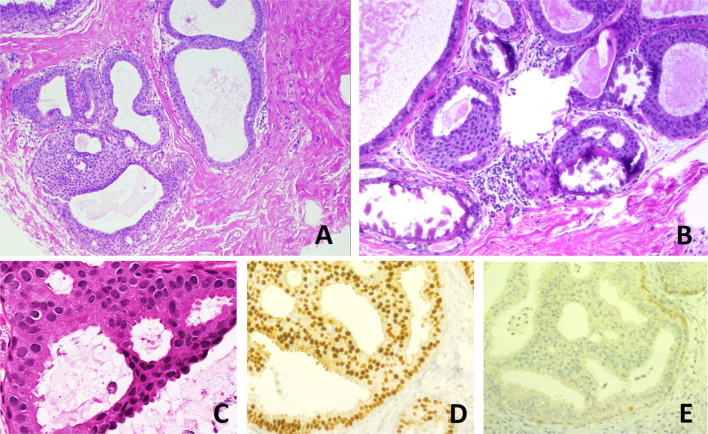
Fig. 1.
Histological appearance of atypical ductal hyperplasia (ADH) and immunohistochemical phenotype. a One focus (< 2 mm) of two architecturally disarranged cross sections of tubuli showing a monotonous intraductal proliferation with secondary intraluminal architecture. Hematoxylin and Eosin stain. b One area of an ADH with associated calcifications intraluminal. Hematoxylin and Eosin stain. c Higher magnification of ADH shows low-grade nuclear atypia and monotonous cell proliferation along with secondary intraluminal architecture. Hematoxylin and Eosin stain. d Strong and uniform expression of estrogen receptors (ER). ER immunohistochemistry. e Lack of basal cytokeratins (CK5/6). CK5/6 immunohistochemistry

